Thursday, December 31, 2009
Our ministry...
I thank the Lord for them...
"From him the whole body, joined and held together by every supporting ligament, grows and builds itself up in love, as each part does its work." -Ephesians 4:16
Our ministry:
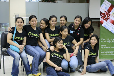
GCF Connect - Call Center Ministry .
Wednesday, December 23, 2009
It is the season...
Behold, a child is born!
"For to us a child is born, to us a son is given, and the government will be on his shoulders. And he will be called Wonderful Counselor, Mighty God, Everlasting Father, Prince of Peace." -Isaiah 9:6
"For I know the plans I have for you," declares the LORD, "plans to prosper you and not to harm you, plans to give you hope and a future. Then you will call upon me and come and pray to me, and I will listen to you. You will seek me and find me when you seek me with all your heart. -Jeremiah 29:11-13
Merry Christmas and a happy new year everyone!
Friday, December 11, 2009
Update: My Ultrasound Mammotome experience
I'm sorry if I failed to update some of you...
Here's what happened-- hindi po natuloy ang medical procedure ko nung December 8, 2009, 2pm sa Medical City.
At sa totoo lang, para na akong sirang plaka kasi ilang beses ko na ito naikwento sa mga tao sa office at sa iba sa church. Hehe. Pero oks lang, tandang-tanda ko naman kasi kung ano nangyari. So here goes...
Nakasalang na ako sa operating room/bed...
- The Surgeon/Doctor told me that I will be given local anesthesia. Ang ikinagulat ko nung sinabi nya na kada bukol tuturukan nya (ouch!) and I guess isa yun sa nagpakaba sa akin.
- First shot of anesthesia: I remember the doctor told me calmly-- "Okay this will sting ha." And it did, pero natiis ko naman. So after the shot, I tried to stay calm. The anesthesia will make me numb naman anyway!
- And now, the time has come for my surgeon to cut me in the area where she will have to place the thing (heck, di ko alam kung ano tawag dun! LOL) na mag-va-vacuum nung bukol na tatanggalin...
***to be continued...
Tuesday, December 1, 2009
Know that the Lord is God!
Just want to share this and ask na rin for your prayers... Hehe.
"Know that the LORD, He is God;
It is He who has made us, and not we ourselves;
We are His people and the sheep of His pasture."
-Psalm 100:3 (New King James Version)
I will undergo ultrasound mammotome breast biopsy (see more info below) next week, December 8, 2009 around 2pm.
Do pray for me and with me.
Salamat sa mga naitext ko na previously about this na patuloy ang pananalangin for me.
I'm confident that I will see more of His goodness. Ü
How a Breast Biopsy using the Mammotome® System Works
Here's what you or someone you care about can expect when undergoing a breast biopsy using the MRI, x-ray, or ultrasound Mammotome® System:
Preparation for the procedure
1. Patient positioning depends on the imaging method that is used. Under ultrasound imaging, a doctor will obtain images of the breast tissue while the patient lies on her back. In stereotactic (x-ray) procedures, the patients lie facedown on a special table. The woman's breast protrudes through a hole in the table's surface, where it is lightly compressed and immobilized while a computer produces detailed images of the abnormality. In the case of MRI imaging, the patient lies face down on a breast coil, and the coil is moved inside a magnet for a computer to acquire images.
2. The patient is given a local anesthetic (injection) to minimize any discomfort.
Biopsy Procedure with the Mammotome® System
3. Once the area to be biopsied has been located and mapped, the Mammotome® probe is inserted through a small ¼-inch incision in the breast.
4. When the probe is positioned at the area of concern, it gently vacuums, cuts and removes tissue samples for examination. The samples are passed through the hollow chamber of the probe into a collection chamber. Because the Mammotome® probe is directional, multiple specimens can be collected without having to remove and reinsert the device.
5. To continue to monitor your breast health following a breast biopsy using the Mammotome® system, the doctor may place a small stainless steel marker or a collagen plug with an embedded titanium marker to identify the biopsy site. While this tiny marker is undetectable to the patient, it enables a physician to keep track of an abnormality in future mammograms.
6. The Mammotome® probe is removed after the samples have been collected, and the single incision is closed with a small adhesive bandage.
(taken from: www.breastbiopsy.com )
Subscribe to:
Posts (Atom)